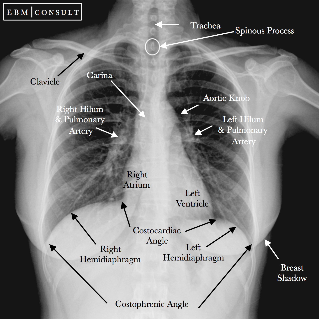
Chest X Ray Clavicle . The posterior shoulder should be in contact with image receptor (ir) or tabletop, without rotation of body. imaging of the clavicle. rotation and heart size. If the patient is rotated to their right, then heart size may be underestimated. The sternum is also included on a frontal view but it overlies other midline structures and so is obscured. The standard ap view of the clavicle is taken with the patient upright or sitting, with arms at the sides, chin raised, and looking straight ahead. If the patient is rotated to their left, then the heart may appear enlarged. the radiographic series of the clavicle is utilized in emergency departments to assess the clavicle,.
from www.ebmconsult.com
The standard ap view of the clavicle is taken with the patient upright or sitting, with arms at the sides, chin raised, and looking straight ahead. the radiographic series of the clavicle is utilized in emergency departments to assess the clavicle,. The posterior shoulder should be in contact with image receptor (ir) or tabletop, without rotation of body. The sternum is also included on a frontal view but it overlies other midline structures and so is obscured. imaging of the clavicle. If the patient is rotated to their right, then heart size may be underestimated. rotation and heart size. If the patient is rotated to their left, then the heart may appear enlarged.
Radiology Chest Xray Normal
Chest X Ray Clavicle The posterior shoulder should be in contact with image receptor (ir) or tabletop, without rotation of body. the radiographic series of the clavicle is utilized in emergency departments to assess the clavicle,. The posterior shoulder should be in contact with image receptor (ir) or tabletop, without rotation of body. If the patient is rotated to their right, then heart size may be underestimated. rotation and heart size. The standard ap view of the clavicle is taken with the patient upright or sitting, with arms at the sides, chin raised, and looking straight ahead. The sternum is also included on a frontal view but it overlies other midline structures and so is obscured. imaging of the clavicle. If the patient is rotated to their left, then the heart may appear enlarged.
From www.ebmconsult.com
Radiology Chest Xray Normal Chest X Ray Clavicle If the patient is rotated to their left, then the heart may appear enlarged. The standard ap view of the clavicle is taken with the patient upright or sitting, with arms at the sides, chin raised, and looking straight ahead. imaging of the clavicle. The posterior shoulder should be in contact with image receptor (ir) or tabletop, without rotation. Chest X Ray Clavicle.
From dxomrrlxr.blob.core.windows.net
What Is A Chest Pa And Lateral X Ray at William Randel blog Chest X Ray Clavicle the radiographic series of the clavicle is utilized in emergency departments to assess the clavicle,. The posterior shoulder should be in contact with image receptor (ir) or tabletop, without rotation of body. The sternum is also included on a frontal view but it overlies other midline structures and so is obscured. imaging of the clavicle. If the patient. Chest X Ray Clavicle.
From www.istockphoto.com
Chest Xray Fractures Left Clavicle Anterior 2nd Rib Posterior Rib 45 Chest X Ray Clavicle The posterior shoulder should be in contact with image receptor (ir) or tabletop, without rotation of body. If the patient is rotated to their left, then the heart may appear enlarged. rotation and heart size. The standard ap view of the clavicle is taken with the patient upright or sitting, with arms at the sides, chin raised, and looking. Chest X Ray Clavicle.
From www.researchgate.net
Sample Chest XRay Measurement in Clavicle Bone. Download Scientific Chest X Ray Clavicle The posterior shoulder should be in contact with image receptor (ir) or tabletop, without rotation of body. The standard ap view of the clavicle is taken with the patient upright or sitting, with arms at the sides, chin raised, and looking straight ahead. the radiographic series of the clavicle is utilized in emergency departments to assess the clavicle,. The. Chest X Ray Clavicle.
From www.cureus.com
Cureus Pathological Clavicle Fracture Initial Presentation of Chest X Ray Clavicle rotation and heart size. The standard ap view of the clavicle is taken with the patient upright or sitting, with arms at the sides, chin raised, and looking straight ahead. the radiographic series of the clavicle is utilized in emergency departments to assess the clavicle,. If the patient is rotated to their left, then the heart may appear. Chest X Ray Clavicle.
From www.shutterstock.com
Chest Xray Fracture Left Clavicle Stock Photo 1568857609 Shutterstock Chest X Ray Clavicle The standard ap view of the clavicle is taken with the patient upright or sitting, with arms at the sides, chin raised, and looking straight ahead. rotation and heart size. imaging of the clavicle. If the patient is rotated to their left, then the heart may appear enlarged. If the patient is rotated to their right, then heart. Chest X Ray Clavicle.
From www.istockphoto.com
Chest Xray Fractures Left Clavicle Anterior 2nd Rib Posterior Rib 45 Chest X Ray Clavicle If the patient is rotated to their left, then the heart may appear enlarged. the radiographic series of the clavicle is utilized in emergency departments to assess the clavicle,. If the patient is rotated to their right, then heart size may be underestimated. The posterior shoulder should be in contact with image receptor (ir) or tabletop, without rotation of. Chest X Ray Clavicle.
From www.dreamstime.com
Chest Xray Film of a Patient with Fracture of Left Clavicle, Ribs 2rd Chest X Ray Clavicle the radiographic series of the clavicle is utilized in emergency departments to assess the clavicle,. The standard ap view of the clavicle is taken with the patient upright or sitting, with arms at the sides, chin raised, and looking straight ahead. rotation and heart size. If the patient is rotated to their left, then the heart may appear. Chest X Ray Clavicle.
From www.researchgate.net
Three radiographs, including (a) chest AP, (b) both clavicle AP, and Chest X Ray Clavicle rotation and heart size. If the patient is rotated to their left, then the heart may appear enlarged. The standard ap view of the clavicle is taken with the patient upright or sitting, with arms at the sides, chin raised, and looking straight ahead. If the patient is rotated to their right, then heart size may be underestimated. The. Chest X Ray Clavicle.
From www.saem.org
Chest Radiograph Chest X Ray Clavicle The sternum is also included on a frontal view but it overlies other midline structures and so is obscured. If the patient is rotated to their left, then the heart may appear enlarged. imaging of the clavicle. the radiographic series of the clavicle is utilized in emergency departments to assess the clavicle,. If the patient is rotated to. Chest X Ray Clavicle.
From www.stepwards.com
Interpreting A Chest XRay Stepwards Chest X Ray Clavicle rotation and heart size. If the patient is rotated to their right, then heart size may be underestimated. The sternum is also included on a frontal view but it overlies other midline structures and so is obscured. The standard ap view of the clavicle is taken with the patient upright or sitting, with arms at the sides, chin raised,. Chest X Ray Clavicle.
From www.dreamstime.com
High Quality Chest Xray and Shoulder and Clavicle Stock Photo Image Chest X Ray Clavicle rotation and heart size. If the patient is rotated to their right, then heart size may be underestimated. The sternum is also included on a frontal view but it overlies other midline structures and so is obscured. the radiographic series of the clavicle is utilized in emergency departments to assess the clavicle,. The standard ap view of the. Chest X Ray Clavicle.
From www.researchgate.net
A AP radiograph of right clavicle on presentation which showed no Chest X Ray Clavicle imaging of the clavicle. The posterior shoulder should be in contact with image receptor (ir) or tabletop, without rotation of body. If the patient is rotated to their right, then heart size may be underestimated. rotation and heart size. If the patient is rotated to their left, then the heart may appear enlarged. The sternum is also included. Chest X Ray Clavicle.
From www.dreamstime.com
Chest Xray Lungs, Heart, Ribs, Clavicle. Medicine Stock Image Chest X Ray Clavicle the radiographic series of the clavicle is utilized in emergency departments to assess the clavicle,. The standard ap view of the clavicle is taken with the patient upright or sitting, with arms at the sides, chin raised, and looking straight ahead. imaging of the clavicle. The sternum is also included on a frontal view but it overlies other. Chest X Ray Clavicle.
From www.alamy.com
Film xray clavicle AP show fracture clavicle bone Stock Photo Alamy Chest X Ray Clavicle The standard ap view of the clavicle is taken with the patient upright or sitting, with arms at the sides, chin raised, and looking straight ahead. If the patient is rotated to their right, then heart size may be underestimated. the radiographic series of the clavicle is utilized in emergency departments to assess the clavicle,. imaging of the. Chest X Ray Clavicle.
From www.dreamstime.com
Chest X Ray with Fractured Clavicle Stock Photo Image of anatomy Chest X Ray Clavicle The sternum is also included on a frontal view but it overlies other midline structures and so is obscured. The standard ap view of the clavicle is taken with the patient upright or sitting, with arms at the sides, chin raised, and looking straight ahead. imaging of the clavicle. If the patient is rotated to their right, then heart. Chest X Ray Clavicle.
From www.dreamstime.com
High Quality Chest Xray and Shoulder and Clavicle Stock Photo Image Chest X Ray Clavicle The sternum is also included on a frontal view but it overlies other midline structures and so is obscured. the radiographic series of the clavicle is utilized in emergency departments to assess the clavicle,. rotation and heart size. If the patient is rotated to their left, then the heart may appear enlarged. The standard ap view of the. Chest X Ray Clavicle.
From www.kenhub.com
Normal chest xray Anatomy tutorial Kenhub Chest X Ray Clavicle rotation and heart size. If the patient is rotated to their right, then heart size may be underestimated. The standard ap view of the clavicle is taken with the patient upright or sitting, with arms at the sides, chin raised, and looking straight ahead. the radiographic series of the clavicle is utilized in emergency departments to assess the. Chest X Ray Clavicle.
